
Magnetic Resonance Imaging (MRI): Cervical Spine
What's an MRI (Magnetic Resonance Imaging)?
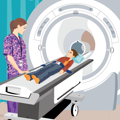
An MRI (magnetic resonance imaging) is a safe and painless test that uses magnets and radio waves to make detailed pictures of the body's organs, muscles, soft tissues, and structures. Unlike a CAT scan, an MRI doesn’t use radiation.
MRIs are done in hospitals and at radiology centers.
What Is a Cervical Spine MRI?
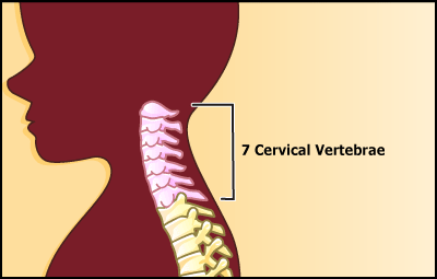
An MRI of the cervical spine produces detailed pictures of the cervical spine (the bones in the back of the neck).
Why Are Cervical Spine MRIs Done?
A cervical (SER-vih-kul) spine MRI can detect a variety of conditions in the neck and upper back area, including problems with the soft tissues within the spinal column, such as the spinal cord, nerves, and disks.
Doctors might order an MRI to evaluate the anatomy of the seven cervical spine bones or spinal cord, or to look for injuries in the area.
A cervical spine MRI also can help doctors:
- Evaluate symptoms such as pain, numbness, tingling or weakness in the arms, shoulders, or neck area.
- Find some types of chronic diseases of the nervous system.
- Diagnose tumors, bleeding, swelling, infections, or inflammatory conditions in the vertebrae or surrounding tissues.
What if I Have Questions?
If you have questions about the cervical spine MRI or the results of the test, speak with your doctor. You can also talk to the MRI technician before the exam.
 Note: All information is for educational purposes only. For specific medical advice,
diagnoses, and treatment, consult your doctor.
Note: All information is for educational purposes only. For specific medical advice,
diagnoses, and treatment, consult your doctor.
© 1995- The Nemours Foundation. KidsHealth® is a registered trademark of The Nemours Foundation. All rights reserved.
Images sourced by The Nemours Foundation and Getty Images.


